Although the placement of chest tubes usually falls to physicians, many healthcare workers have had to care for patients with chest tubes in place, either in a hospital setting or during transport. Chest tubes are a critical factor in certain respiratory emergencies, so let’s review the indications for placement and some of the dangerous complications you can avoid when caring for such patients.
Fundamentals of Chest Tubes
Chest tubes are flexible plastic tubes that are inserted into the pleural space through a small incision made in the side of the chest. They are used to evacuate air, fluid, pus, or blood from the chest cavity. They are primarily inserted to treat traumatic injuries, most typically:
- Pneumothorax – air escaping into the pleural space, either from a disruption in the lung tissue or from an opening in the chest wall, resulting in a collapsed lung
- Hemothorax – bleeding into the pleural space, which prevents the lungs from inflating fully and can lead to collapse
- Hemopneumothorax – a combination of the two conditions above
Once the necessary equipment is assembled, a small incision is made on the affected side of the body, above the fifth rib at the midaxillary line. The tube is inserted into the pleural space and sutured in place. Air and fluid can then flow from the chest or be drawn out through the application of suction. A radiograph of the chest is usually taken to ensure proper placement.
This simple life-saving procedure is highly effective in reestablishing the negative pressure within the chest and reinflating collapsed lungs. But there can be complications, especially during transport. They include:
- Accidental removal
- Recurrent pneumothoraces
- Broken collection chambers
- Parenchymal injuries
- Infection
Protect Your Patient
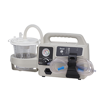
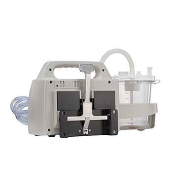
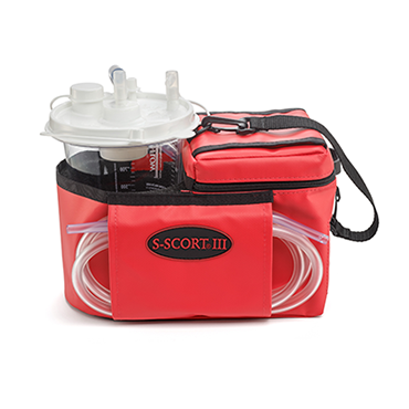
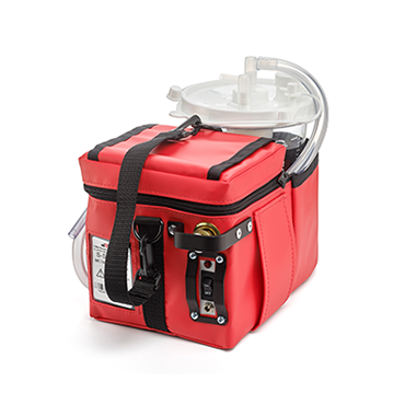
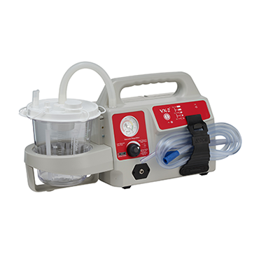
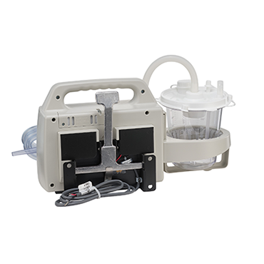
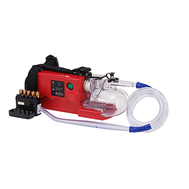
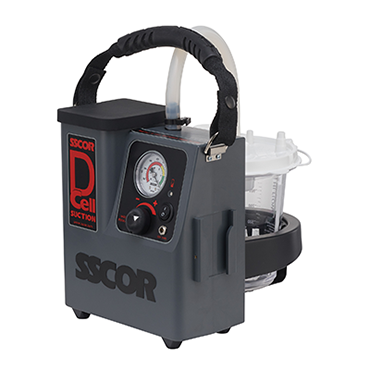
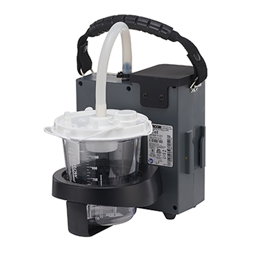
You can prevent these complications by protecting the patient during inter- and intra-facility transports. A portable suction device will be needed for patient relocation if the chest tube is connected to suction. Before transporting the patient using a portable suction unit, a few things to check are:
- Be sure the batteries are charged.
- Ensure the unit is clean and operational—turn it on to test it!
- Have extra batteries on hand for lengthy transports.
Once your suction unit is ready, be sure the patient is, too. Here are a few reminders:
- Ensure that all connections are secure, either using tape or wire banding.
- Make sure the bandage over the incision is securely taped and occlusive.
- Mark the depth of the tube using a felt-tip marker and continually monitor during transport.
- If a drainage unit is used, be sure to keep it below the level of the chest. (Always use an appropriate chest drain system. Chest tubes should never be attached directly to wall or portable suction.)
The chest drain system properly regulates the vacuum level, prevents backflow, and collects fluids. Always use an appropriate chest drain system. Chest tubes should never be attached directly to wall or portable suction. The chest drain system properly regulates vacuum level, prevents backflow, and collects fluids.
- Coil tubing to prevent kinks.
- Document any output in the collection chamber, as well as the type of fluid.
- Do NOT clamp the tube for transport because this will likely cause a tension pneumothorax.
Although chest tubes can correct life-threatening injuries, they must be monitored carefully, especially during patient transport. Be sure to follow these guidelines to ensure your patient’s safety.
Editor's Note: This blog was originally published in October 2021. It has been re-published with additional up-to-date content.


