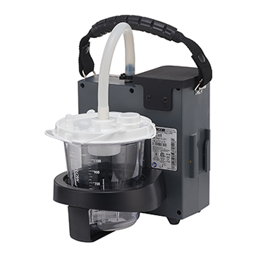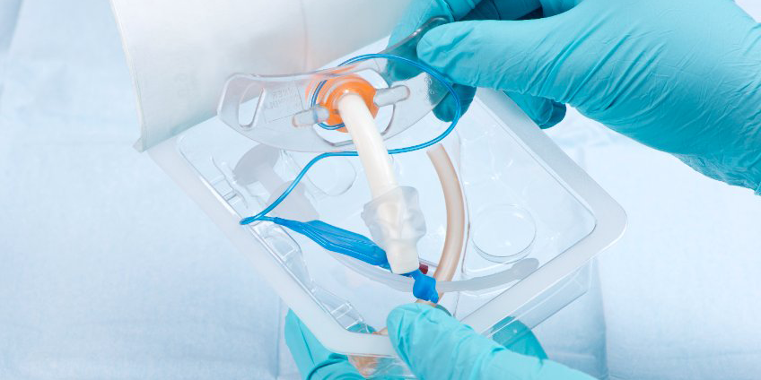
Many people, from infants and children to adults with certain medical conditions, rely on tracheostomies for breathing assistance. A tracheostomy — which involves placing a tube through the front of the neck into the windpipe (trachea) to provide an air passage — is necessary in many cases, but it can also come with various risks and airway management concerns for patients and their providers.
To help you and your team provide the best care to patients undergoing tracheostomies, we’ve put together some information about when tracheostomies are needed, the risks and how providers can better prepare for possible airway emergencies resulting from tracheostomies.
What is a tracheostomy and when is it needed?
A tracheostomy, also known as a tracheotomy, may be temporary or permanent depending on the severity of a person’s condition and medical needs. There are various instances in which patients might require tracheostomies to assist with breathing. Although this procedure is often related to health problems that require the long-term use of a ventilator, below are some other scenarios that might require tracheostomies:
- Sudden blocking of the airway after a traumatic face or neck injury
- Paralysis, neurological problems or other conditions that make it harder to cough up secretions from the throat and thus require direct suctioning of the windpipe (trachea) to clear the airway
- Preparation for a major head or neck surgery to assist with breathing during recovery
- Severe trauma to the head or neck that obstructs breathing
Though less common than with adults, doctors may sometimes recommend tracheostomies for children if they need help breathing, their airway is blocked or too small, they need help removing extra mucus or they’re experiencing aspiration.
Possible airway emergencies
Any procedure that involves the breaking of skin can carry tremendous risks, and tracheostomies are no exception. There are several possible airway emergencies that providers should remain on high alert for when treating tracheostomy patients.
Some of these include:
Blocked or dislodged tubes: A blocked or dislodged tube can cause substantial breathing problems. The blocking or dislodging of a tube may occur for several reasons, like simple movements of the head, neck or body or moving and rolling of the patient by medical staff and caregivers. Factors that may increase the risk of dislodgement include:
- Treating larger or obese patients
- Patients with thick or short necks
- Low tracheostomy placement
- High patient movement
- Loose tracheal ties
- Traction on ventilatory tubing
- Use of positive-pressure ventilation
Trapped air in the tissue or under the skin of the neck (subcutaneous emphysema): Trapped air during tracheostomies most often occurs in the skin covering the chest or neck, and it can cause breathing problems and damage to the trachea or esophagus. Subcutaneous emphysema may result from various traumatic injuries, such as a facial bone fracture, collapsed lung and a rupture or tear in the airway, esophagus or gastrointestinal tract.
Some methods providers can employ to effectively treat this condition include offering airway or breathing support using oxygen from an external delivery device or endotracheal intubation, using a chest tube, conducting blood tests and administering fluids through a vein.
Lung collapse: Lung collapse occurs when air escapes outside of the lung into the chest, and pressure prevents the lung from expanding properly. Some identifiable symptoms of lung collapse include:
- A sudden sharp, stabbing pain in the chest
- Rapid breathing or shortness of breath
- Rapid heart rate
- Low blood pressure
- Patient turning blue
- Lung expansion on one side
- Anxiety and/or fatigue
The long-term impacts of lung collapse vary. If only a small amount of air is trapped in the chest, there might not be additional complications, but if the volume of air is large or it impacts the heart, the outcome can be life-threatening.
Emergency preparation and training
All providers should be trained in emergency scenarios for treating tracheostomy patients. Emergency simulations are good opportunities to practice responding quickly and effectively in different tracheostomy situations, such as tracheostomy changes, inner cannula changes, mechanical ventilation and alarm scenarios, suctioning and other tracheostomy care.
In addition to running emergency simulations, there are various safety protocols providers must practice when preparing to treat tracheostomy patients:
- Always watch for signs of respiratory distress.
- Pre-hyperoxygenate the patient if required and according to your agency’s policy.
- If you remove oxygen while performing tracheostomy care, remember to replace it often to reoxygenate the patient.
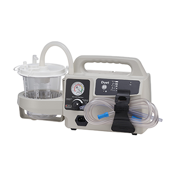
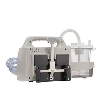
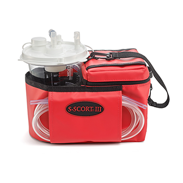
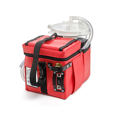
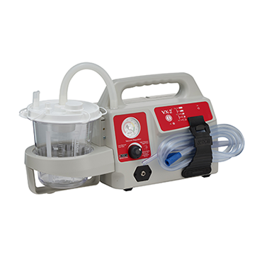
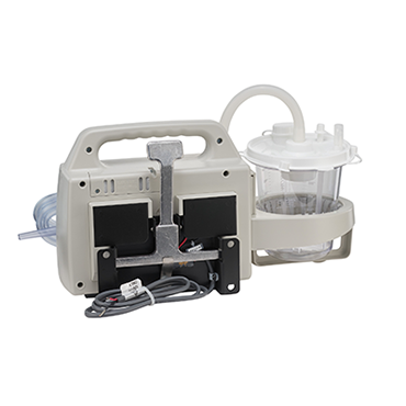
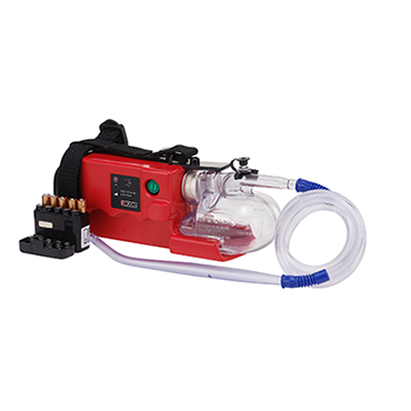
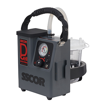
Having the right equipment on-hand is vital and equipping yourself with a variety of catheters and suction devices will help you remain prepared for a range of emergency patient scenarios. Providers should always keep flexible suction tubing and equipment at the bedside when treating tracheostomy patients, and different catheter sizes should be available during treatment.
Looking for more information on how to prepare for airway emergencies? Read SSCOR’s breakdown of different catheter tips and equipment sizes to ensure you and your team are ready for any emergencies your tracheostomy patients face.


