Medical providers have long used pulse oximeters as a quick and easy way to assess blood oxygen levels. However, the amount of CO2 a person expires is an equally useful piece of information that provides key details about ventilation. Capnography provides clear data about the amount of CO2 expired at each stage of respiration. Using capnography during medical suction can reduce the risk of hypoxia and provide additional details about patients at risk of serious suction-related complications.
Capnography: The Basics
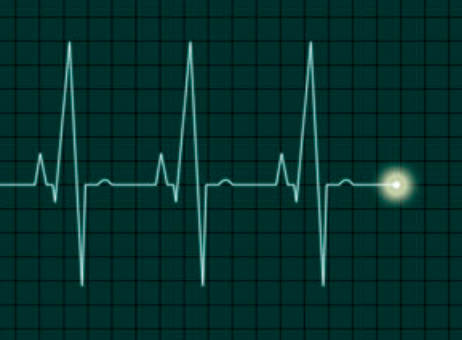
Waveform capnography measures how much CO2 a patient expires at each stage of respiration in real time. The readout provides two pieces of data: a number and a graph. The number is the amount of CO2 pressure at the end of each exhalation – the normal range is 35-45 mmHg. This figure is called end-tidal CO2, or ETCO2.
The graph is a wave that shows CO2 levels during the entirety of the respiration cycle. A healthy graph is a rectangular shape that goes up and down at regular intervals. Other shapes—particularly a shark fin shape that goes sharply up and down—indicate poor ventilation. Capnography can measure CO2 using nasal prongs or an adapter attached to a BVM.
Capnography measures that which pulse oximetry and patient observation often cannot. A patient may have good oxygenation readings because they can inhale sufficient oxygen, even when they are headed toward respiratory failure. Capnography offers an early warning of poor ventilation and can help guide treatment decisions. For example, a patient with high ETCO2 at risk of respiratory arrest may need BVM ventilation.
What ETCO2 Reveals About the Patient
Though suction is generally safe and potentially life-saving, all medical procedures present some risks. Suctioning is no exception, particularly in a patient with serious respiratory health issues such as pneumonia or COPD.
When ETCO2 is higher than normal and is accompanied by sharp points on a capnography graph, respiratory failure is the most likely culprit. High ETCO2 happens when increased effort does not efficiently eliminate CO2. This can lead to respiratory arrest or respiratory failure. Yet some patients have normal pulse oximetry readings because they are still able to inhale enough oxygen.
When ETCO2 is low, this may indicate perfusion, psychological distress due to an anxiety attack, or a metabolic issue. A pulmonary embolism can cause a decrease in CO2-rich blood returning to the lungs, while diabetic ketoacidosis may lower ETCO2 and elevate respiration. Here again, it’s clear that capnography adds data points that can support rapid response and an accurate diagnosis.
Benefits of Capnography
Capnography offers real-time feedback that can help with patient assessment and treatment decisions. A shift to a rectangular pattern on a capnographic readout suggests a patient is responding well to suction or other breathing treatments. Other benefits include:
- The ability to measure lung ventilation rather than just blood oxygenation.
- Information about the amount of respiratory effort a patient is undertaking, not just the results of that effort.
- A reduced risk of hyperventilation—a common and preventable side effect of BVM ventilation.
- The ability to predict respiratory problems that pulse oximeters may fail to predict.
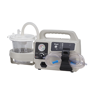
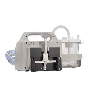
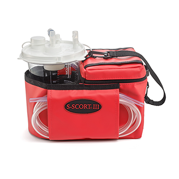
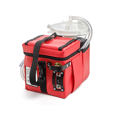
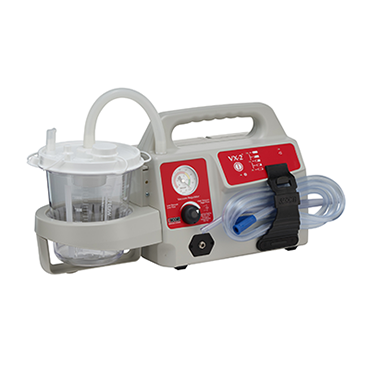
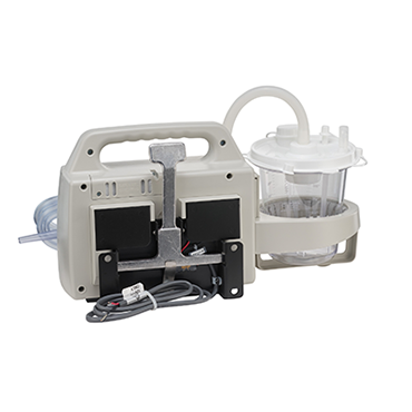
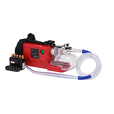
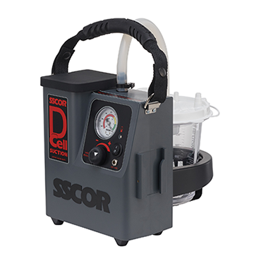
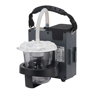
Capnography works best alongside other quality equipment, including a reliable suction unit. When a patient suffers respiratory distress, an airway obstruction, or airway trauma, seconds count. Portable emergency suction machines allow you to quickly tend to the patient, without the risk and effort of moving them to an ambulance. For help selecting the right portable emergency suction machine for your agency, download our free e-book, The Ultimate Guide to Purchasing a Portable Emergency Suction Device.
Editor's Note: This blog was originally published in July, 2019. It has been re-published with additional up to date content.


