The stats speak for themselves. Respiratory distress is what sends 10% of children to emergency departments. Additionally, one in seven seniors has a lung disease. Between 1980-2014, more than 4.6 million American adults died from chronic respiratory diseases.
Performing comprehensive respiratory assessments can detect problems before they become emergencies. Additionally, in hypoxic patients or those with airway obstructions, a respiratory assessment provides important information about the patient’s status and clues about next treatment steps.
Let’s look at the basics of performing an effective and comprehensive respiratory assessment.
Patient History
A respiratory assessment must begin with a detailed patient history. Ask about previous respiratory illnesses, chronic respiratory conditions, and cardiovascular health. If the patient has an infection or is in respiratory distress, get as many details as possible about the event preceding the emergency. Ask about the patient’s vaccine history, as well.
This is also an ideal chance to determine whether the patient has special needs that might affect the assessment. Preterm infants, for example, have weaker respiratory muscles than children and adults, while infants and young children have a more rapid rate of respiration. Ensure you know what’s normal for the patient population you serve, as well as the specific patient you are treating.
Observation
Observe the patient for important respiratory clues:
- Check the rate of respiration.
- Look for abnormalities in the shape of the patient’s chest.
- Ask about shortness of breath and watch for signs of labored breathing.
- Check the patient’s pulse and blood pressure.
- Assess oxygen saturation. If it is below 90 percent, the patient likely needs oxygen.
In infants and newborns:
- Check for flaring nostrils, which could indicate breathing problems.
- Look for retractions or bulging of the muscles between the ribs, which suggest difficulty getting enough air.
Auscultation
Hearing the sounds of the patient breathing provides vital information about the patient’s overall health. Auscultate the chest, back, and sides with a focus on signs of loud or labored breathing. Signs of abnormal breathing include:
- Crackling, popping, or bubbling sounds, which may indicate pneumonia or pulmonary edema.
- Wheezing, which can signal pulmonary disease, asthma, allergies, or an infection.
- Pleural friction. This grating sound occurs when the pleural surfaces rub together and suggests pneumonia.
Physical Examination
A hands-on exam is critical for detecting abnormalities that simple observation and auscultation cannot. To examine the patient:
- Palpate the back at the tenth rib, positioning a thumb on each rib as the patient breathes deeply. Patients with decreased lung expansion may have a tumor or pneumonia on one side. Poor lung expansion could also indicate pneumothorax.
- Evaluate the thorax by positioning the palms over the thorax and feeling for bulging, tenderness, and retractions while breathing. Feel the ribs for lumps, scars, and swelling.
- Have the patient fold their arms across their chest. Then position both palms on either side of the back, touching the patient’s back with your fingers while the patient says a sentence.
- You should feel buzzing as the patient speaks. If there is fluid in the lungs or a lower respiratory obstruction, the vibrating will be intense because of the ability of fluid to more effectively transmit sound.
Percussion
Percussion can provide additional information about respiratory status. Use the middle or index finger of your dominant hand to tap the areas between each rib through the chest or back. Avoid touching the skin with your other fingers, since this can cause vibrations that compromise the assessment.
Sounds to monitor for include:
- A short and high-pitched or very dull sound over muscle or bone. This suggests respiratory consolidation.
- A loud, long, low-pitched and hollow sound over the lungs or stomach that may suggest bronchitis.
- A dull, thudding sound over large organs such as the liver. This may also be a sign of consolidation.
- A loud, low-pitched sound over the stomach that can indicate pneumothorax or emphysema.
- A high-pitched drum sound is heard when the chest is expanded. This suggests excess air, often due to a collapsed lung.
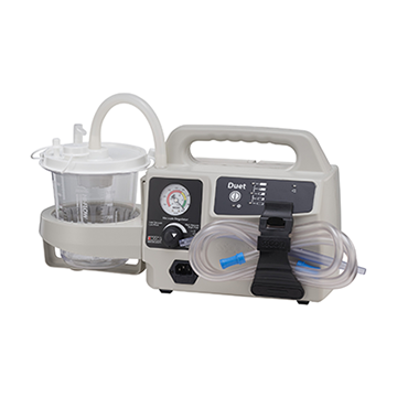
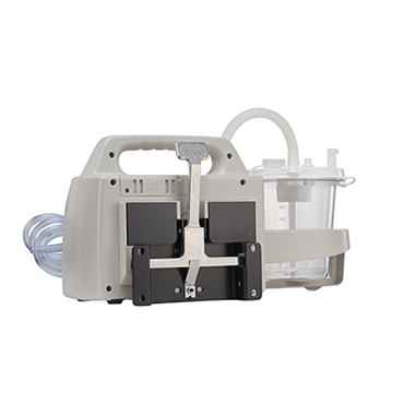
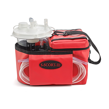
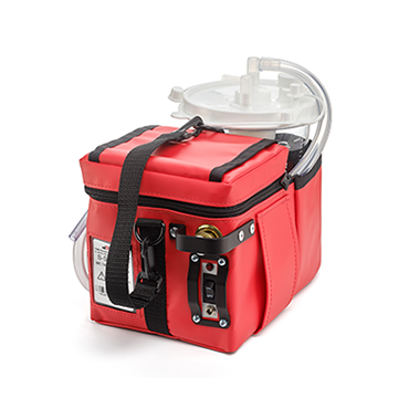
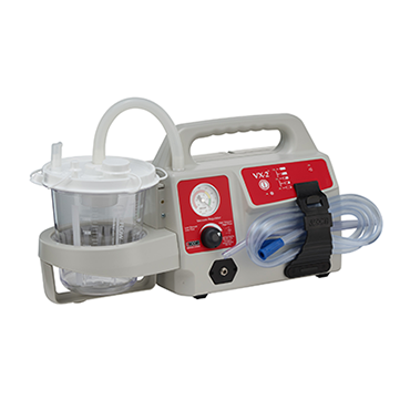
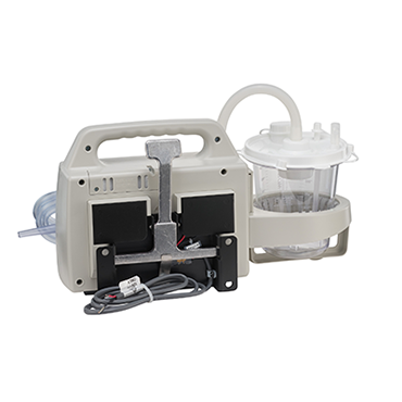
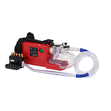
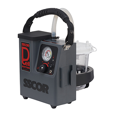
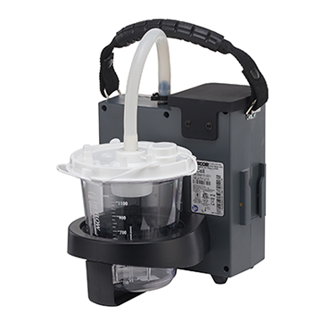
A respiratory assessment provides important details about treatment, and the right treatment may include clearing the airway of obstructions. For help selecting the right equipment for your agency, download our free guide, The Ultimate Guide to Purchasing a Portable Emergency Suction Device.
Editor's Note: This blog was originally published in December 2018. It has been re-published with additional up to date content.


